Cardiac muscle fibers cells also are extensively branched and are connected to one another at their ends by intercalated discs. An intercalated disc allows the cardiac muscle cells to contract in a wave-like pattern so that the heart can work as a pump. Figure 10.21 Cardiac Muscle Tissue Cardiac muscle tissue is only found in the heart. LM × 1600.
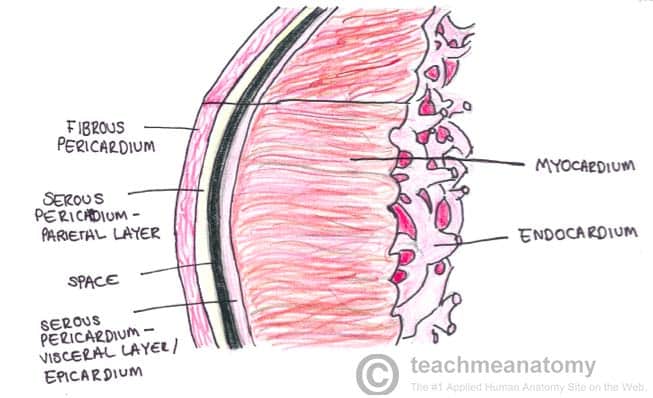
The Pericardium – TeachMeAnatomy
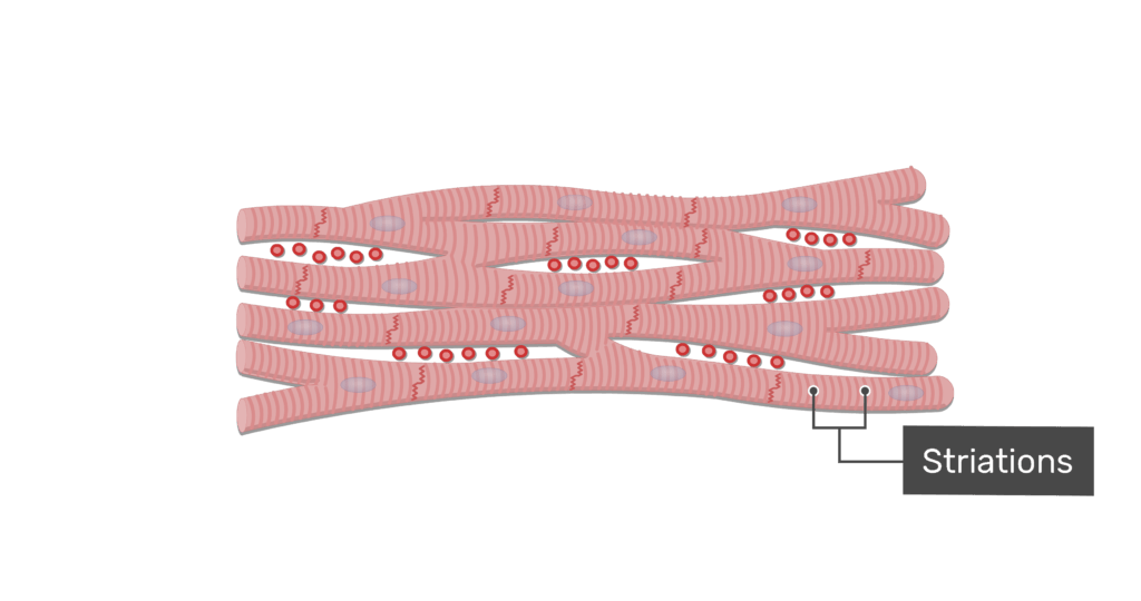
Cardiac Muscle Cardiac muscle tissue occurs only in the heart, where it con-stitutes the bulk of the heart walls. Like skeletal muscle cells, cardiac muscle cells are striated, but cardiac muscle is not volun-tary. Indeed, it can and does contract without being stimulated by the nervous system. Most of us have no conscious control
Source Image: twinkl.co.uk
Download Image
Nov 23, 2022Cardiac muscle fibers are long, branched cells, shaped like cylinders joined end-to-end, with one or two nuclei located centrally. The fibers are separated by collagenous tissue that supports the capillary network of cardiac tissue. The myofilaments of cardiac muscle are arranged in a similar pattern to skeletal muscle, resulting in cross

Source Image: science.org
Download Image
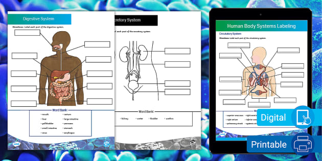
A&P 1- CHAPTER 9 MASTERING ASSIGNMENTS Flashcards | Quizlet
Art-labeling Activity: Figure 18.11 Part A Drag the appropriate labels to their respective targets. Chapter 18 Multiple Choice Question 31 Part A Which is most responsible for the synchronized contraction of cardiac muscle tissue? Correct Chapter 18 Multiple Choice Question 16 Part A If the length of the absolute refractory period in cardiac muscle cells was the same as it is for skeletal

Source Image: pubs.acs.org
Download Image
Art-Labeling Activity Structure Of A Cardiac Muscle Fiber
Art-labeling Activity: Figure 18.11 Part A Drag the appropriate labels to their respective targets. Chapter 18 Multiple Choice Question 31 Part A Which is most responsible for the synchronized contraction of cardiac muscle tissue? Correct Chapter 18 Multiple Choice Question 16 Part A If the length of the absolute refractory period in cardiac muscle cells was the same as it is for skeletal
Terms in this set (9) Identify tissue type 1. Identify tissue type 2. Identify tissue type 3. Study with Quizlet and memorize flashcards containing terms like Skeletal Muscle, Cardiac Muscle, Smooth Muscle and more.
A Complete Workflow for High Throughput Human Single Skeletal Muscle Fiber Proteomics | Journal of the American Society for Mass Spectrometry
Study with Quizlet and memorize flashcards containing terms like The endomysium __________., Art-labeling Activity: The Structure of a Sarcomere, Art-labeling Activity: The structure of a skeletal muscle fiber and more.
how to draw cardiac muscle fibre | how to draw labelled diagram of cardiac muscle fibre – YouTube

Source Image: youtube.com
Download Image
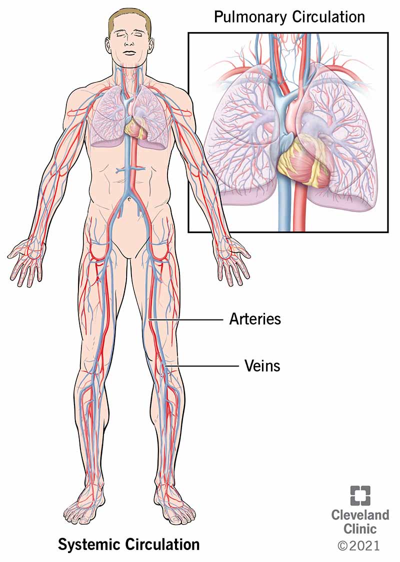
How Your Cardiovascular System Works
Study with Quizlet and memorize flashcards containing terms like The endomysium __________., Art-labeling Activity: The Structure of a Sarcomere, Art-labeling Activity: The structure of a skeletal muscle fiber and more.
Source Image: my.clevelandclinic.org
Download Image
The Pericardium – TeachMeAnatomy
Cardiac muscle fibers cells also are extensively branched and are connected to one another at their ends by intercalated discs. An intercalated disc allows the cardiac muscle cells to contract in a wave-like pattern so that the heart can work as a pump. Figure 10.21 Cardiac Muscle Tissue Cardiac muscle tissue is only found in the heart. LM × 1600.
Source Image: teachmeanatomy.info
Download Image
A&P 1- CHAPTER 9 MASTERING ASSIGNMENTS Flashcards | Quizlet
Nov 23, 2022Cardiac muscle fibers are long, branched cells, shaped like cylinders joined end-to-end, with one or two nuclei located centrally. The fibers are separated by collagenous tissue that supports the capillary network of cardiac tissue. The myofilaments of cardiac muscle are arranged in a similar pattern to skeletal muscle, resulting in cross

Source Image: quizlet.com
Download Image
how to draw diagram of cardiac muscle step by step for beginners ! – YouTube
There are two major types of cardiac muscle cells: myocardial contractile cells and myocardial conducting cells. The myocardial contractile cells constitute the bulk (99 percent) of the cells in the atria and ventricles. Contractile cells conduct impulses and are responsible for contractions that pump blood through the body.

Source Image: m.youtube.com
Download Image
Cardiac muscle tissue: function and labeled diagram | GetBodySmart
Art-labeling Activity: Figure 18.11 Part A Drag the appropriate labels to their respective targets. Chapter 18 Multiple Choice Question 31 Part A Which is most responsible for the synchronized contraction of cardiac muscle tissue? Correct Chapter 18 Multiple Choice Question 16 Part A If the length of the absolute refractory period in cardiac muscle cells was the same as it is for skeletal
Source Image: getbodysmart.com
Download Image
art-labeling activity structure of a skeletal muscle fiber – brainly.com
Terms in this set (9) Identify tissue type 1. Identify tissue type 2. Identify tissue type 3. Study with Quizlet and memorize flashcards containing terms like Skeletal Muscle, Cardiac Muscle, Smooth Muscle and more.

Source Image: brainly.com
Download Image
How Your Cardiovascular System Works
art-labeling activity structure of a skeletal muscle fiber – brainly.com
Cardiac Muscle Cardiac muscle tissue occurs only in the heart, where it con-stitutes the bulk of the heart walls. Like skeletal muscle cells, cardiac muscle cells are striated, but cardiac muscle is not volun-tary. Indeed, it can and does contract without being stimulated by the nervous system. Most of us have no conscious control
A&P 1- CHAPTER 9 MASTERING ASSIGNMENTS Flashcards | Quizlet Cardiac muscle tissue: function and labeled diagram | GetBodySmart
There are two major types of cardiac muscle cells: myocardial contractile cells and myocardial conducting cells. The myocardial contractile cells constitute the bulk (99 percent) of the cells in the atria and ventricles. Contractile cells conduct impulses and are responsible for contractions that pump blood through the body.
